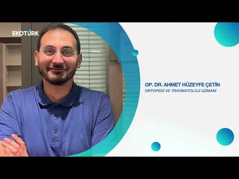
Duygu Bal | Op.Dr. Ahmet Huzeyfe Çetin | Hanse Nur Özyurt | Erhan Özgür Our Job is Health
Duygu Bal | Op.Dr. Ahmet Huzeyfe Çetin | Hanse Nur Özyurt | Erhan Özgür İşimiz Sağlık 📞 444 3 515 numarasını …

Aile Hastanesi Urology Department provides diagnosis and treatment services for urological cancer surgeries, paediatric urology surgeries and male sexual dysfunctions in addition to routine operations such as prostate and bladder TUR surgeries, prostate HOLEP open and closed stone surgeries (ureteroscopy, flexible ureterorenoscopy, percutaneous nephrolithotomy).
In the treatment of urinary disorders due to prostate enlargement (BPH), medication and surgical treatment methods (TUR, prostate surgery with Holmium Laser -HoLEP) are applied. In addition, intensive studies are carried out in areas related to prostate health such as prostate cancer screening and treatment of prostate infections.
UROONCOLOGY
The department performs nerve-sparing radical prostatectomy (for prostate cancer) and radical cystectomy (for bladder cancer). Other urological oncology operations such as kidney and testicular cancer are also performed with the support of anaesthesiology and intensive care services.
ENDOUROLOGY AND STONE DISEASES
In the Urology Department of Aile Hospital, endoscopic (closed system) surgeries are also performed in the treatment of urinary system stone disease.
Urology department provides services in endourological treatment of stone disease (ureteroscopy, RIRS (Flexible Ureterorenoscopy, percutaneous kidney stone surgery - PCNL).
FEMALE UROLOGY
Diagnosis and treatment services are provided for diseases such as urinary incontinence (stress and urge incontinence) and interstitial cystitis.
ANDROLOGY
Examination, examination and treatment for erectile dysfunction called erectile dysfunction and ejaculation problems in men and male infertility conditions related to male infertility are planned and carried out together with other medical specialities.
PRP, stem cell therapy, ESWT and penile prosthesis applications are performed in erection problems. Penis lengthening and thickening surgeries are performed in penis aesthetics.
PAEDIATRIC UROLOGY
Paediatric urology deals with congenital or acquired urinary and genital system diseases that can be seen in the newborn and later periods, starting from the womb, and their treatment. Examples of these diseases include hydrocele and cord cyst, undescended testicle, circumcision, emergency conditions of testicles and bags (acute scrotum and testicular torsion), vesicoureteral reflux and hypospadias.
UROLOGICAL DISEASES AND TREATMENT METHODS
PROSTATE ENLARGEMENT - BPH
It occurs in 1/3 of men over the age of 50. It is not a malignant tumour. This growth is thought to occur with hormonal effect. Benign growth of the prostate should not be confused with cancer. The mechanisms of formation are different, and after one occurs, the other one is not a continuation of it. However, both can be found together in 15 per cent of cases.
After the prostate enlarges, it compresses the urinary duct with pressure, the patient urinates with difficulty and even becomes unable to urinate in time.
SYMPTOMS
At first, the diameter of urine decreases and urine flow slows down. When urinating while standing or sitting, the patient cannot urinate forwards. Urine comes intermittently. Urine flows in drops. There is a feeling of incomplete emptying. Urine does not come immediately and waits for a while. Since urination slows down, the duration of urination is prolonged. Frequent urination occurs. Normally, there is no urination at night or urination may occur only once.
As in every patient, the patient’s complaints and additional diseases are evaluated in detail. Prostate enlargement is not the only cause of lower urinary system voiding complaints. It should not be forgotten that diseases that disrupt bladder contraction, diabetes, some neurological diseases, urinary tract stenosis, infections can also cause similar complaints.
Therefore, patients should be evaluated in detail before treatment.
DIAGNOSIS OF PROSTATE ENLARGEMENT
Urinalysis, detailed evaluation of the bladder, prostate and kidneys by ultrasonography, urine flow measurement by voiding test, PSA blood test are the main tests used. The urine remaining in the bladder after voiding is measured by ultrasound.
In addition, the International Prostate Symptom Score (IPSS), in which patients evaluate themselves, helps us to evaluate the severity of complaints.
In some cases, it may be necessary to examine the urethra and bladder endoscopically (with a camera).
WHEN IS SURGERY NECESSARY?
Patients who do not benefit from medication or do not use medication
Those whose quality of life and social life are adversely affected by urinary complaints
Patients who cannot urinate and are catheterised
Patients with urinary tract inflammation
Patients who are unable to urinate and whose bladder or kidney begins to deteriorate due to residual urine
Patients with stones in the bladder
Patients with enlarged prostate and bleeding in the urine
The above-mentioned conditions are patients who absolutely need surgery. Each patient should be evaluated individually. Many factors such as existing comorbidities, medications, general health status, expectations from surgery, sexual life, social-work life are taken into consideration and the most appropriate treatment method is selected for the patient.
PROSTATE OPERATIONS
TUR-P: It is a procedure in which prostate tissue, especially smaller than 80-100 grams, is cut with the help of a wire with electric current by entering through the urinary canal with a camera device. Bipolar and plasmacokinetic techniques using different electric currents are widely used today. It is based on cleaning the prostate tissue blocking the urinary canal.
HoLEP: It is the complete removal of enlarged prostate tissue using laser energy. Its advantages compared to the tour method are much less bleeding. It can be applied to prostate of any size. It is a candidate treatment option to be the gold standard method. It is the most suitable surgical treatment option especially for large prostates. Does not impair erection. Probe duration and hospital stay are short. No semen discharge after sexual intercourse
Open prostatectomy: It is the removal of enlarged (over 100 g) prostate tissue with or without an abdominal incision into the bladder. There is a risk of bleeding and may require prolonged hospitalisation. The duration of the catheter is longer. Return to normal life requires time.
WHAT IS HOLEP?
It is the process of enucleating the prostate tissue using Holmium laser energy, that is, completely removing it from the capsule. It has been applied since 1995. Especially in recent years, the popularity of the technique has increased in parallel with the developments in laser technology. Especially thanks to high-energy laser devices, the surgery is performed more successfully and with fewer complications.
HOW IS HOLEP PERFORMED?
It is performed under general or spinal anaesthesia (numbing below the waist). The penis is entered through the external urinary canal with special tools with a camera. It is examined whether there is a stenosis in the urinary tract. The enlarged prostate tissue is seen. The bladder is entered and the bladder is examined for tumours and stones. Then, the prostate is scratched with a laser at certain points and the grey-white capsule tissue surrounding the prostate tissue is reached. The vessels spreading from the capsule into the prostate are easily sealed with the laser and bleeding is much less.
Depending on the size or shape of the prostate tissue, it is removed from the shell in 2-3 or single pieces and thrown into the bladder. The prostate tissue thrown into the bladder is broken up with another instrument (morcellator) and vacuumed out of the body. After all the pieces are removed, final controls are performed and the procedure is terminated by inserting a catheter.
The removed prostate tissue is sent to pathology to investigate the presence of cancer.
WHO IS HOLEP RECOMMENDED FOR?
HoLEP is a surgical method that can be applied to everyone recommended for prostate surgery. Since the HoLEP technique has some advantages over other surgical methods, it is also applied more safely in cases where other methods cannot be applied.
Although the classical TUR method is not recommended for prostates larger than 80-100 g, HoLEP can be applied much more successfully especially for large prostates. In addition, there is no upper limit on prostate size for HoLEP. HoLEP is also successfully performed in the same session in patients with bladder stones accompanying prostate size.
It can also be applied more safely than other techniques in patients with cardiovascular diseases, coronary stents or by-pass in the past, and patients who use blood thinners due to vascular occlusion.
HOW MANY DAYS TO STAY IN HOSPITAL AFTER HOLEP?
Generally, the patient is discharged from hospital on the 2nd or 3rd day after the operation.
DOES HOLEP AFFECT SEXUAL FUNCTIONS?
In benign prostate enlargement surgeries such as HoLEP, the prostate is scraped from the capsule. When compared in terms of the energy used, holmium laser energy has been shown to affect the prostate capsule only to a depth of 0.4 mm. In other words, the energy used to separate the prostate tissue does not damage the nerves that provide hardening. The electrical energy used in TURP surgery can spread to a depth of 4 mm.
The common disadvantage of HoLEP and other TURP-open prostatectomy techniques in terms of sexual functions is that they cause retrograde ejaculation or inability to ejaculate. Since the prostate tissue is completely cleaned, the semen goes backwards to the bladder at the moment of orgasm and is then excreted in the urine. This is not harmful for health.
As a result, HoLEP will not damage the erection but will cause retrograde ejaculation during orgasm.
PROSTATE CANCER
Although there are many risk factors, prostate cancer mainly develops in men due to exposure of the prostate gland to male hormone (testosterone) with aging. It is recommended that every man over the age of 40 if there is a family history of prostate cancer and over the age of 50 if there is no family history of prostate cancer should have an annual examination and PSA test in the blood. Prostate health index, multiparametric prostate MRI and advanced genetic examinations are performed in suspicious cases. MRI-USG (Magnetic resonance imaging-ultrasound) fusion biopsy procedure is performed rectally or perineally under sedation for patients for whom biopsy is decided. Thanks to this method, which is routinely applied in our clinic, cancers that cannot be detected by ultrasound become visible with the help of MR and allow targeted sampling of suspicious images. In the surgical treatment of prostate cancer, Laparoscopic Radical Prostatectomy and Open Radical Prostatectomy operations are successfully performed in our hospital. Taking into account the risk factors of the disease, the need for pelvic lymph node dissection and additional treatments according to the pathology results are decided by your urologist.
KIDNEY AND URETER STONES
WHAT ARE THE DAMAGES OF KIDNEY STONES?
Kidney stones, which cause severe agonising pain, can cause kidney obstruction, loss of kidney function and kidney failure if appropriate treatment is not given in time. In fact, the infection that occurs can be mixed into the bloodstream and cause disruption of the functions of other organs and severe infections (urosepsis).
While the disease is seen with different frequencies in different geographies, the prevalence in our country is 11%. In other words, approximately one in every 9 people in our country has stone disease. It is 2-3 times more common in men than women. It is especially common between the ages of 40-60.
HOW DOES KIDNEY STONE GIVE SYMPTOMS?
The most common symptoms are periodic increasing and decreasing pain (renal colic), burning and discolouration in the urine. The pain is blunt, severe, writhing and passing. Patients usually describe the pain as the most severe pain they have ever experienced. It is even likened to labour pain experienced by women.
When the stones fall from the kidney into the urinary canal, the pain is in the groin area and may radiate downwards. The pain does not increase or decrease with position. Nausea, vomiting, burning and discolouration of urine may also accompany the pain. There may also be fever if there is an accompanying infection. If the kidney obstruction is bilateral, there is a decrease in urine output.
Stone patients with no complaints are also rarely encountered.
HOW IS KIDNEY STONE DIAGNOSED?
We use biochemical and radiological methods to make a diagnosis. For this, we refer patients to the laboratory and radiology departments:
The first of the tests is urinalysis. In the urine of stone patients, blood cells, crystals and bacteria are seen if there is an infection. As a blood test, kidney function tests (creatinine, urea, BUN) should be performed first. If infection is suspected, complete blood count and infection markers should be performed.
The definitive diagnosis of stone disease is made by radiological methods.
Ultrasound: It is the first preferred method. It is the most important advantage of not using radiation and is the first preferred method in children and pregnant patients.
X-ray: Since approximately 70% of the stones contain calcium, they can be visualised by X-ray (direct urinary system radiography). Stones that can be visualised by X-ray are called opaque (Ca-phosphate, Ca-oxalate) and those that cannot be visualised are called radiolucent or non-opaque stones (uric acid, ammonium urate, xanthine, drug-induced stones). Some stones cannot be visualised clearly and are called semi-opaque (magnesium-ammonium-phosphate: struvite, apatite and cystine).
Tomography: With its introduction, computed tomography (CT) is now recognised as the most sensitive (99%) method for diagnosing stone patients. Its biggest advantages are that it provides information about the location, size and hardness (density) of the stone and the relationship of the kidney with the intra-abdominal organs. Although radiation exposure is the biggest disadvantage, this concern has been eliminated with the use of low dose radiation emitting devices.
Other:
Nuclear methods (DMSA, DTPA, MAG3) may be used in cases of loss of renal function or structural disorders leading to impaired drainage. Magnetic resonance imaging (MRI) is also the preferred method of examination in pregnant patients in whom X-ray methods are inconvenient.
HOW IS KIDNEY STONE TREATED?
The treatment of kidney stones is planned according to the location and size of the stone and the patient’s clinical condition and concomitant diseases.
Stone crushing with extracorporeal sound waves (ESWL)
It is a treatment method that does not require anaesthesia and is applied to small kidney stones. This method is based on the principle of transmitting sound waves produced from a generator through the skin to the kidney and the stone and breaking the stones. Several sessions are required for complete stone-free treatment. The broken stone fragments are spontaneously excreted from the body through urine. It is not preferred for pregnant women and patients taking blood thinners.
The hardness of the stone, the distance between the stone and the skin, the point where the stone is located in the kidney, and the size of the stone are factors affecting the success. Taking all these factors into consideration, it can be performed safely and successfully in suitable patients.
Uretero-renoscopic lithotripsy (URS-RIRS)
It is known as bloodless and incisionless surgery. Under anaesthesia, the stones in the ureter or kidney are broken down by entering the urinary tract (urethra) first into the bladder (bladder) and then into the urinary canal (ureter) with instruments (endoscopes) that have a camera at the very thin end and allow us to see inside the body. The stones in the upper ureter and kidney are pulverised by using a flexible endoscope called ureterorenoscope and laser and left to fall out spontaneously. Larger stone fragments are removed with instruments called basket. It can be safely applied in patients taking blood thinners. Its biggest advantages are that it is performed without any incision in the body and short hospital stay. The disadvantage is its low success rate in large kidney stones and the need for several sessions.
Percutaneous nephrolithotomy (PNL)
PNL is a procedure in which a tube-pathway extending from the skin to the kidney is created on the back under anaesthesia and the stones are broken and removed by entering the kidney with endoscopes (nephroscope) through this pathway. It is the most successful method especially in large kidney stones (larger than 2cm) where other methods fail.
Apart from these methods, laparoscopic and open surgery methods can also be applied in stone treatment. Open surgery is almost abandoned today and is preferred in limited patient groups where other methods fail. Laparoscopic surgery is applied as an alternative method to open surgery.
Edited by: DOÇ.DR. MEHMET NURİ BODAKÇİ

Duygu Bal | Op.Dr. Ahmet Huzeyfe Çetin | Hanse Nur Özyurt | Erhan Özgür İşimiz Sağlık 📞 444 3 515 numarasını …

Duygu Bal | Op.Dr. Ahmet Huzeyfe Çetin | Hanse Nur Özyurt | Erhan Özgür İşimiz Sağlık 📞 444 3 515 numarasını …
